
A 70-year-old patient, operated on after an unstable pelvic fracture, remains bedridden for three weeks as a precaution. The result: significant muscle wasting, calf thrombosis, and an extended rehabilitation period of several months. This scenario often repeats itself because managing recovery after a pelvic fracture relies on a delicate balance between rest and resuming movement.
Early mobilization after pelvic fracture: the trap of prolonged immobility
We spontaneously associate fractures with strict rest. For the pelvis, this logic can backfire on the patient. Orthopedic rehabilitation recommendations lean towards early mobilization as soon as the fracture’s stability allows. In some post-surgical protocols, standing is encouraged as early as the day after the procedure.
Staying lying down for too long leads to cascading complications. Muscle wasting affects the stabilizing muscles of the pelvis (glutes, psoas, transverse abdominis) within a few days. The thromboembolic risk significantly increases with immobility. In older adults, the loss of autonomy sets in quickly and is corrected slowly.
The practical question is not “should we move?”, but “when and how?”. Partial weight-bearing with a walker, mobilization exercises while lying down, respiratory work: these simple actions reduce the overall recovery time. To better understand the recovery and duration of immobilization after a pelvic fracture, it is essential to distinguish stable fractures, which tolerate a quick return, from unstable fractures, which require a more cautious schedule but never total immobility.

Rehabilitation mistakes that delay pelvic healing
Three mistakes frequently occur in practice, and they often accumulate in the same patient.
Resuming full weight-bearing too quickly
The opposite of excessive immobility also exists. Some patients, relieved to be able to stand, force full weight-bearing before bone consolidation. The pelvis bears the weight of the trunk: premature weight-bearing can displace a partially consolidated fracture site. The surgeon or rehabilitation physician establishes a progressive weight-bearing schedule (touch weight, partial weight, full weight) that should not be accelerated independently.
Neglecting pain as a warning signal
Pain during rehabilitation is not just a discomfort to overcome. Sharp pain while walking, discomfort that increases over sessions, nighttime awakenings related to the fractured area: these signs should trigger a radiographic check. Pushing through pain delays healing instead of accelerating it.
Forgetting overall muscle work
Rehabilitation after a pelvic fracture is not limited to the injured area. The muscles of the posterior chain, deep abdominals, and hip stabilizers form a functional unit. Focusing solely on joint mobility without muscle strengthening exposes patients to postural compensations that generate secondary pain in the back or knees.
Nutrition and complications: two underestimated angles during recovery
There is much talk about physiotherapy, but rarely about what happens outside of sessions. Bone consolidation directly depends on nutritional intake.
- Foods rich in calcium and vitamin D (dairy products, fatty fish, green vegetables) support the formation of bone callus. A deficiency measurably slows healing.
- Proteins are the building material for muscle reconstruction. After a period of immobilization, even a short one, protein demand increases.
- Sufficient hydration and preventing constipation (common with opioid analgesics) avoid discomfort that limits mobilization efforts.
Regarding complications, pelvic fractures are notable for their proximity to pelvic organs and blood vessels. Urinary sequelae, chronic sacral pain, and persistent walking disorders affect a significant proportion of patients. Regular medical follow-up after radiological consolidation remains necessary, as functional sequelae can sometimes appear weeks after “bone healing.”
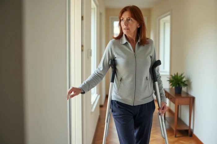
Home modifications and fall prevention during healing
Returning home is a critical phase. It is observed that many relapses or slowdowns in recovery stem from the daily environment, not from the rehabilitation program itself.
- Removing rugs and floor obstacles reduces the risk of falls, the leading cause of refracture in older patients.
- Installing grab bars in the bathroom and toilet allows for standing and sitting without excessively straining the pelvis.
- Using a toilet seat riser and a shower stool prevents deep hip flexions, which are painful and potentially dangerous during the consolidation phase.
- Ensuring a bed at the appropriate height: lying down and getting up from a bed that is too low heavily engages the pelvic muscles.
These modifications are not mere comfort details. They condition the patient’s ability to follow their progressive mobilization program without incident. Hospitals are increasingly incorporating home assessments before the patient’s return, especially for operated unstable fractures.
The duration of rehabilitation after a pelvic fracture varies according to the severity and general condition of the patient. Bone consolidation generally takes a few months, but functional rehabilitation can extend well beyond that. Returns vary on this point, depending on age, type of fracture, and quality of follow-up. Respecting each step of the protocol without rushing through the stages remains the best way to regain lasting mobility.